Ultrasound™
Gaumard Ultrasound™
A high-fidelity, portable simulator designed to immerse trainees in realistic, scenario-based training and build clinical skills that translate to real-world care.
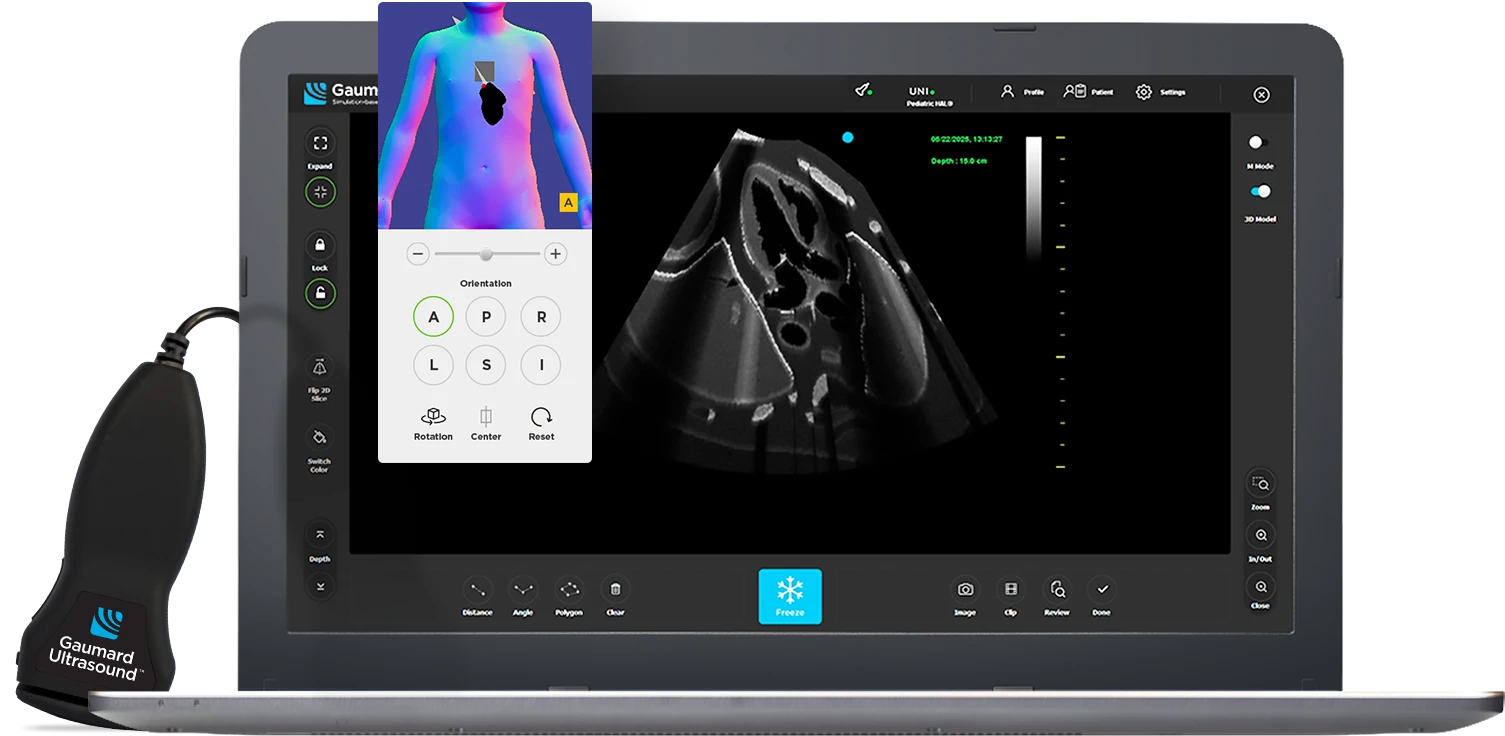
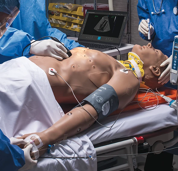
Gaumard Ultrasound™ simulates the core functions of an ultrasound machine, with natural transducer motion and lifelike imaging that reinforce proper technique and diagnostic confidence. Gaumard Ultrasound is the first and only ultrasound simulator to offer full, unrestricted scanning across the anterior torso on advanced patient simulators, giving trainees the freedom to scan as they would in actual clinical settings.
Purpose-Built for Teaching Clinical Thinking

Dynamic scenario control.
Powered by UNI® 3.
Seamlessly integrated with our intuitive simulation software, allowing you to modify pathologies, adjust vitals, and challenge trainees on the fly-all within a single interface. It’s responsive training that evolves with every decision, giving instructors complete control over the learning environment.
Pause. Teach. Debrief.
Instructional tools designed to pause and enhance learning. Lock the scan to hold the image while the anatomy continues to evolve; ideal for highlighting changes, reinforcing clinical reasoning, or guiding group instruction. Capture key frames, record video, add annotations, and walk through performance during debrief.
Visualize in 3D.
Reinforce anatomical accuracy.
The interactive model alongside the ultrasound view can help connect 2D imaging with 3D anatomy in real time. Visualizing probe position and orientation within the body clarifies structure, depth, and spatial relationships, accelerating clinical comprehension.
Build technique.
Teach precision.
Use real-time ultrasound controls to reinforce scanning accuracy and clinical decision-making. Adjust depth, zoom, and contrast, freeze images to pause for instruction, use on-screen measurement and annotation tools—distance, angle, polygon—to validate findings and teach precision.
Real-time ultrasound.
Real-world accuracy.
Responsive imaging that reacts instantly to probe manipulation, just like in clinical practice. Trainees get immediate visual feedback, reinforcing proper scanning techniques and promoting muscle memory for better performance under pressure.
A transducer that moves like the real thing.
Includes a physical probe with unrestricted, natural range of motion across the anterior torso—from the base of the neck to the lower abdomen. There are no fixed imaging windows or “points”. Trainees can scan freely, building spatial awareness and motor coordination critical for real-world ultrasound use.
Preloaded with pathology. Ready for any case.
From pneumothorax and hemothorax to liver lacerations, the system offers a broad library of adult or pediatric cases. Run focused assessments or full-code trauma simulations.
Physiology and imaging, fully in sync.
Ultrasound output is synchronized with the patient simulator’s vital signs, so what trainees see on screen matches what they monitor clinically. Heart rate, respiratory rate, and visible motion in the lungs or myocardium all align, reinforcing situational awareness and deepening diagnostic accuracy.
Designed for POCUS and eFAST.
Support for the most critical diagnostic protocols used in trauma and emergency care. Whether you're training prehospital providers or critical care teams, trainees build skills that translate directly to high-acuity environments.
- Pediatric and adult compatible: Compatible with both HAL® S5301 and Pediatric HAL® S2225, the system expands your simulation capabilities without requiring separate hardware or workflows.
- Complete the picture with vitals: Add the optional Gaumard Vitals™ Monitor to overlay real-time physiological data. Participants must synthesize ultrasound results with changing vitals, just like they would at the bedside.
- Built to integrate: No patchwork required. Designed to work seamlessly within the Gaumard ecosystem. No third-party software, no custom installations, and no steep learning curves. Just plug in, connect, and start training.
- Tailored to your training style: Display modes, scanning options (B-Mode and M-Mode), and a customizable interface allow you to adjust settings for every training scenario. It’s flexibility that adapts to your curriculum, not the other way around.
- Healthy
- Left Pneumothorax
- Left Hemothorax
- Left Pleural Effusion
- Right Pneumothorax
- Right Hemothorax
- Right Pleural Effusion
- Splenic Hematoma
- Splenic Rupture with Blood in Rectovesical Pouch
- Liver Laceration
- Pericardial Effusion
- Pericardial Tamponade with Right Atrial Compression
- Pneumoperitoneum
- Appendicitis
- Bladder Rupture
- Cholecystitis
- Healthy
- Left Pneumothorax
- Left Hemothorax
- Right Pneumothorax
- Right Hemothorax
- Splenic Hematoma
- Splenic Rupture with Blood in Rectovesical Pouch
- Liver Laceration
- Pericardial Effusion
- Pneumoperitoneum
- Appendicitis
- Bladder Rupture
Gaumard Ultrasound™
Package Contents


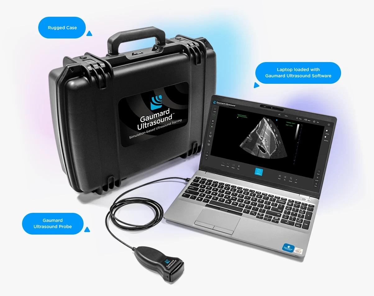
For HAL® S5301
For Pediatric HAL® S2225
Patent pending
Tech Support
Connect to a Gaumard
Representative
Download User Guides
Get the current User Guides
for your products
Gaumard Software
Download the latest software
and updates
Bridging the gap between theory and practice.
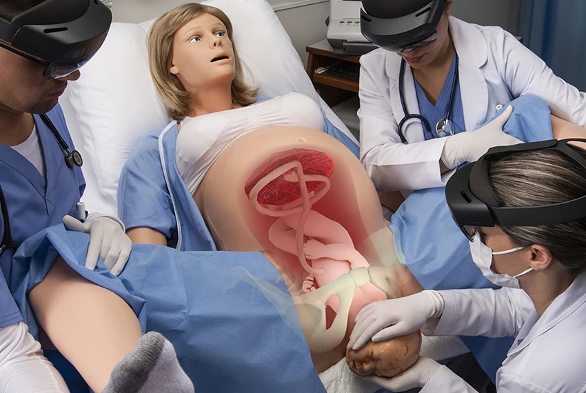
By synchronizing holograms with the physical world, Obstetric MR allows learners to see inside VICTORIA and observe the dynamic physiology underlying difficult deliveries to promote deeper learning. Wireless connectivity supports up to 6 participants, with each observing their unique vantage point.
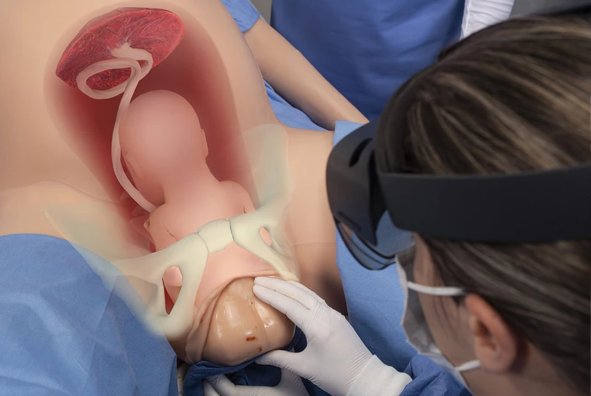
Seamless and powerful scenario integration.
Obstetric MR adds a new layer of learning to normal vaginal birth, shoulder dystocia, and breech scenarios. It integrates seamlessly with VICTORIA’s new Simulation Learning Experiences childbirth scenarios as well as your custom scenarios and on-the-fly operation.
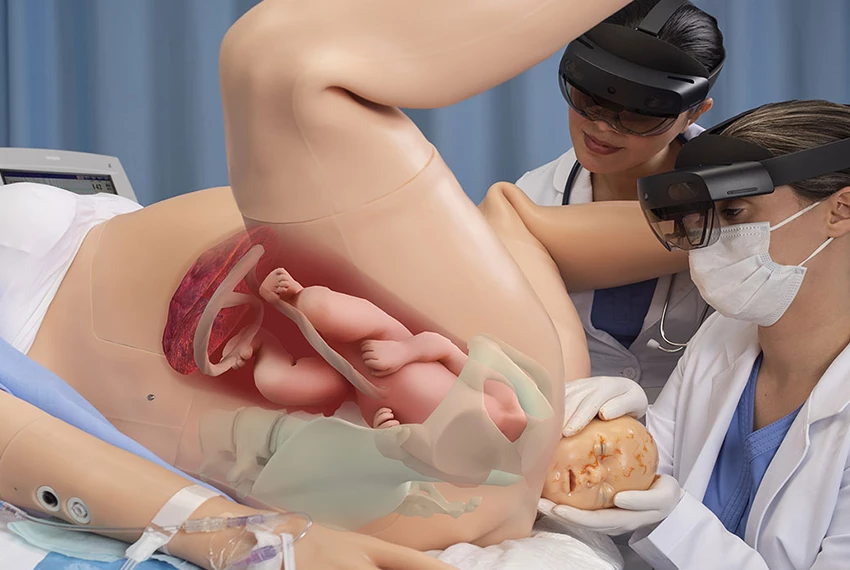
Observe clinical intervention in real-time.
Obstetric MR, VICTORIA’s internal network of sensors, and the powerful UNI® control software work together to provide learners with real-time visual feedback. Learners can study the rotation of the pelvis and the fetal shoulder during McRoberts and suprapubic pressure maneuvers as they perform them. It is an entirely new way to observe and understand clinical cause and effect through hands-on practice.

